Heart Disease: Knowing Different Types and How to Prevent It

Introduction

Heart disease, often known as cardiovascular disease, is one of the main causes of mortality globally. Cardiovascular disorders pose a major danger to world health, accounting for 17.9 million deaths annually. This article discusses the many forms of heart disease and offers advice on how to avoid it.

Learning about cardiovascular disease

Heart disease, also referred to as cardiovascular disease, is a group of disorders that impact the anatomy and operation of the heart. Among these disorders include arrhythmias, valvular heart disease, coronary artery disease, and heart failure. It is crucial to remember that every kind of cardiac disease has unique traits and consequences.

The cardiovascular system, which is made up of the heart and blood arteries, is responsible for the circulation of blood. Heart disease, which is characterised by blood artery constriction or blockage, frequently affects this system. The majority of heart attacks and strokes are brought on by underlying cardiac conditions, and they might occur suddenly. Heart disease symptoms might vary, but they frequently include discomfort in different body areas, dyspnea, and chest pain.

Types of Heart Disease

The most prevalent kind of heart illness, Coronary Artery illness (CAD), affects millions of people globally. Atherosclerosis, or the accumulation of plaque in the arteries that feed blood to the heart muscle, causes the arteries to narrow and stiffen, which leads to coronary artery disease (CAD). This may lessen the heart’s blood supply, which might result in a heart attack or chest discomfort.

Heart Failure:

This condition is brought on by the heart’s inability to pump blood as effectively as it should. Heart failure is characterised by a heart that still functions, but with less force than usual. Diabetes, high blood pressure, and coronary artery disease are among the causes of heart failure.

Arrhythmias:

Heart rhythm abnormalities are known as arrhythmias. The heart may beat abnormally, too quickly, or too slowly. Arrhythmias can be benign or indicative of a more serious cardiac problem, and they might feel like a racing or fluttering heart.

One of the four heart valves may be damaged or defective in valvular heart disease. The heart’s blood flow is controlled by these valves. Blood flow through the heart can be hampered by conditions like stenosis, or narrowing of the valve, and regurgitation, or leakage of the valve.

Other Types:

Heart disease can also manifest as congenital heart defects, which are abnormalities of the heart present from birth, cardiomyopathies, which are disorders of the heart muscle, and rheumatic heart disease, which is heart damage brought on by rheumatic fever.

Every kind of cardiac disease is different and needs a different approach to therapy. Early identification, the right medical intervention, and lifestyle modifications are frequently crucial to the management of many disorders.

Hazards Associated with Heart Disease

• It is essential to comprehend the risk factors for both management and prevention. These risk factors fall into two categories: those that are changeable and those that are not.

Changeable Risk Elements

Lifestyle Decisions: The risk of heart disease is greatly increased by an unhealthy diet, physical inactivity, tobacco use, and excessive alcohol intake. These habits can raise blood pressure, cholesterol, and glucose levels. They can also cause overweight and obesity.

Medical Conditions:

Heart disease is significantly influenced by a number of conditions, including high blood pressure, high cholesterol, diabetes, and obesity.

Non-Adaptable Risk Elements:

Age: As people age, their risk of cardiac issues rises.

Gender: Risk is higher for men in general, but it also rises for women after menopause.

Genetics and Family History: Having a family history of heart disease raises an individual’s risk.

A thorough discussion of these risk factors aids in the comprehension of the significance of lifestyle decisions and the requirement for routine medical exams by readers.

Preventive Techniques

A multifaceted strategy is needed to prevent heart disease, including dietary adjustments, frequent tests, and, in certain situations, medicines.

Healthy Diet:

Stress the value of eating a well-balanced diet that is high in whole grains, fruits, vegetables, and lean meats. It’s critical to cut back on salt, trans fats, and saturated fats.

Frequent Physical Activity:

List the advantages of frequent exercise, including lowered blood pressure, better weight control, and heart health improvements.

Reducing Alcohol and Tobacco Use:

Talk about the harmful consequences that smoking and binge drinking have on heart health.

Frequent Health screens:

Suggested for readers are routine check-ups that involve screens for diabetes, cholesterol, and blood pressure.

Stress management:

Draw attention to the negative effects of stress on heart health and offer practical solutions like yoga, meditation, or therapy.

Medication:

To manage problems like high blood pressure or cholesterol, some people must take drugs. Emphasize how important it is to take prescription drugs as directed.

Having Heart Disease and Getting By

Living with heart disease entails controlling the illness with medicine, lifestyle modifications, and routine medical attention.

Medication and Treatment Compliance:

Stress how crucial it is to heed medical advice and take prescribed drugs on a regular basis.

Lifestyle Modifications:

Talk about how people with heart disease should alter their behaviors, food, and level of activity.

Tracking and Handling Symptoms:

Provide instruction on identifying symptoms and knowing when to get medical attention.

Psychological Impact and help Systems:

Admit that having cardiac disease has an emotional and psychological toll. Suggest that people seek out help from mental health counselors, support groups, or medical experts.

Rehabilitation Programs:

Describe how cardiac rehabilitation programs help people live better lives generally and with better hearts.

Case Study: Cardiovascular Disease Prevention at the Source

A 53-year-old Caucasian gentleman with a complicated medical history is the subject of a real-life case study that illustrates the management and prevention of cardiovascular disease. The example emphasizes the significance of tailored care in avoiding heart disease. This patient had a lengthy history of smoking, pre-diabetes, class I obesity, and a family history of early coronary artery disease (CAD). His cholesterol and blood pressure readings were alarming, and his low-density lipoprotein (LDL) level of 141 mg/dL suggested an increased risk of heart disease.

Assessing the patient’s 10-year risk for atherosclerotic cardiovascular disease (ASCVD), which was 16.3% and classified as intermediate risk, was the main preventive method. His lifetime ASCVD risk was also estimated to be 50%. Starting moderate-intensity statin medication was advised due to his high LDL-C values, risk-enhancing variables such as metabolic syndrome, and a family history of early ASCVD. Medication called a statin is useful in preventing heart disease because it lowers cholesterol.

The control of his hypertension was a crucial component of his situation. Given his high risk of ASCVD, it was advised that, with a blood pressure of 145/60, he aims for a blood pressure of less than 130/80 mm Hg.
This would entail changing one’s lifestyle in addition to perhaps starting antihypertensive medication therapy with ACE inhibitors, calcium channel blockers, and thiazide diuretics.

Lifestyle therapy was also very important for his control. It was highly advised to stop smoking because smoking has a major negative influence on heart health. A healthy diet, consistent exercise, and weight control were all essential components of his preventative approach.

Conclusion

Summarize the main ideas about the various forms of heart disease, the risk factors associated with them, and the preventative measures that may be taken. Stress the value of taking the initiative to maintain heart-healthy practices, and urge readers to speak with medical experts for individualized guidance and routine examinations. Finish with a message of empowerment and optimism, emphasizing how much the risk of heart disease may be decreased with the correct information and lifestyle choices.

One of the top cardiologists in New York, Dr. Ellen Mellow, provides individualized cardiac and preventive care options. She has over 24 years of expertise in internal medicine and cardiology, and she is dedicated to giving each patient individualized, high-quality treatment. Her work is centered on holistic approaches to heart health, placing equal emphasis on cutting-edge technology and a thorough comprehension of each patient’s unique circumstances. For those in the New York region looking for heart health treatment, Dr. Ellen Mellow is a great resource due to her experience and patient-centered approach. To obtain further details, kindly visit her website at

What Does An Artery Blocked or Narrowed Mean to Your Heart Health?

Introduction

Millions of people around the world are affected by heart health. A leading cause of death in the United States is cardiovascular diseases, so it can’t be overstated how important this issue is. In today’s fast-paced world, the health of our heart is a concern that often lurks in the background, unnoticed until a critical moment. Among the various threats to heart health, These conditions, known medically as atherosclerosis, are not just isolated medical anomalies; they represent a complex interplay between our lifestyle choices, genetic predispositions, and the relentless march of time.

Understanding Arteries and Their Role in Heart Health

The arteries play a pivotal role in our cardiovascular system and overall heart health. They are blood vessels that distribute oxygen-rich blood from the heart to all parts of the body. This process is crucial for delivering the oxygen and nutrients required for the functioning of organs and tissues​​​​.

Types of Arteries and Their Functions:

There are two primary types of arteries, each serving a distinct purpose:

1. Elastic Arteries: These are located close to the heart and have more elastic tissue. Examples include the aorta and the pulmonary artery. They function akin to a football player catching a ball, absorbing the force from the throw, which in this case is the blood pumped from the heart.

Muscular Arteries: These have smooth muscles and are responsible for transporting blood to the body’s tissues, much like a football player running down the field with the ball. They include arteries such as the femoral, radial, and brachial arteries​​.

Anatomy and Structure

Arteries are tubular structures with thick, muscular walls capable of handling high blood pressure from each heartbeat. They don’t need valves as the force of the blood ensures unidirectional flow. The aorta, the largest artery, has a diameter ranging from 10 millimeters to 25 millimeters. The arteries then branch into smaller vessels, reaching the entire body​​​​.

Arterial Supply to Organs

Each organ or body part receives blood supply through specific arteries. For instance, the coronary arteries supply the heart, the carotid arteries provide blood to the brain, head, face, and neck, while the femoral artery caters to the legs​​.

Arteries and Blood Pressure Regulation

Arteries play a crucial role in maintaining blood pressure and controlling blood flow. They adjust by tightening or loosening their muscular walls. Approximately 10% of the body’s blood is in the arteries at any given time​​.

The Exception: Pulmonary Artery

The pulmonary artery is unique as it is the only artery that carries deoxygenated blood from the heart to the lungs for oxygenation​​.

Heart Disease Statistics in the United States

The leading cause of death in the U.S. is cardiovascular disease (CVD), with 928,741 deaths reported in 2020. Between 2018 and 2019, CVD costs were staggering at $407.3 billion in direct and indirect costs. A significant percentage of CVD-related deaths in 2020 were caused by coronary heart disease, followed by strokes and other cardiovascular conditions.

Heart disease prevention relies heavily on maintaining arterial health, as this data illustrates. Understanding the function and significance of arteries in the cardiovascular system is fundamental for managing and preventing heart-related conditions effectively.

Blocked and Narrowed Arteries: Causes, Effects, and Complications

Causes of Arterial Blockage and Narrowing

The accumulation of lipids, cholesterol, and other materials on and inside the artery walls is known as atherosclerosis, and it is the primary cause of clogged and constricted arteries. The accumulation of plaque can restrict the arteries and obstruct blood flow.

In some instances, plaque may rupture, leading to a blood clot. Notably, atherosclerosis is a progressive disease and can affect arteries throughout the body​​.

Key factors contributing to the development of atherosclerosis include:

• High blood pressure: Damages the inner layer of an artery.

• High cholesterol and triglycerides: Contribute to plaque buildup.

• Smoking and tobacco use: Cause damage to the arterial walls.

• Diabetes and insulin resistance: Increase the risk of arterial damage.

• Obesity: Associated with higher cholesterol levels and inflammation.

• Inflammation: From diseases like arthritis, lupus, psoriasis, or inflammatory bowel disease.

• Lifestyle factors: Such as an unhealthy diet, lack of exercise, and sleep apnea​​.

Symptoms and Diagnosis of Blocked and Narrowed Arteries Symptoms

The symptoms of atherosclerosis, the primary cause of blocked and narrowed arteries, can vary based on the severity and location of the artery affected:

1. Mild Atherosclerosis: Often asymptomatic.
2. Moderate to Severe Atherosclerosis: Symptoms emerge when an artery is significantly narrowed or clogged, restricting blood flow. This can lead to:

• Heart Arteries: Chest pain or pressure (angina).

• Arteries Leading to the Brain: Sudden numbness or weakness in limbs, difficulty speaking, temporary loss of vision, or drooping muscles in the face, signaling a transient ischemic attack (TIA) which can lead to a stroke if untreated.

• Arteries in Arms and Legs: Symptoms of peripheral artery disease, like leg pain during walking or decreased blood pressure in a limb.

• Arteries Leading to Kidneys: High blood pressure or kidney failure​​.

Diagnosis

Diagnosing atherosclerosis involves a combination of physical examination, medical history, and a series of tests:

1. Physical Examination and History: A healthcare provider will perform a physical exam, including listening to arteries with a stethoscope for abnormal sounds (bruits), and inquire about personal and family health history.

2. Diagnostic Tests: Depending on the physical exam results, one or more of the following tests may be recommended:

• Blood Tests: To check blood sugar and cholesterol levels; high levels increase the risk of atherosclerosis. A C-reactive protein test may be done for inflammation.• Electrocardiogram (ECG or EKG): Measures the heart’s electrical activity to determine if there’s reduced blood flow.

• Exercise Stress Test: Used if symptoms occur during exercise, showing heart problems that might be missed otherwise.

• ED: When performing percutaneous coronary intervention (PCI), the quantitative flow ratio (QFR)

• shows the heart’s blood flow using sound waves.

• Doppler Ultrasound: Measures blood pressure at various points to assess blood flow speed in arteries.

• Ankle-Brachial Index (ABI): Compares blood pressure in the ankle and arm to check for atherosclerosis in the leg and foot arteries.

• Cardiac Catheterization and Angiogram: Shows if coronary arteries are narrowed or blocked.

• Coronary Calcium Scan: Uses CT imaging to detect calcium deposits in coronary arteries, indicating a higher risk of heart attack.

• Other Imaging Tests: Like Magnetic Resonance Angiography (MRA) or Positron Emission Tomography (PET) to study arteries for hardening, narrowing, and aneurysms​​.

Latest Advancements in Treatment for Blocked and Narrowed Arteries

When performing percutaneous coronary intervention (PCI), the quantitative flow ratio (QFR)

• A novel technique called the quantitative flow ratio (QFR) is significantly improving outcomes post-PCI. This method, which involves 3D artery reconstruction and measurement of blood flow velocity, offers precise measurements of pressure drop across a blockage. QFR allows doctors to make more informed decisions about which arteries to stent during PCI, leading to improved patient outcomes.

• A study involving 3,825 participants in China found that QFR-guided PCI resulted in better patient outcomes compared to standard angiography-guided procedures. The QFR approach reduced unnecessary stent placements and identified more obstructive lesions that required treatment. This led to lower rates of heart attacks and the need for additional PCI, marking a 35% improvement over standard procedures​​.

Nanoparticle-Based Plaque Removal

• Researchers at Missouri S&T are developing a cellular-level approach to removing arterial plaque using nanoparticles. These nanoparticles, designed to deliver plaque-busting drugs directly to specific cells in the arteries, have shown promise in significantly reducing plaque buildup.

• The new method targets plaque cells and inhibits the pathway for cholesterol and lipid accumulation in the arteries. It also has the potential to remove existing cholesterol from plaque cells, a capability not offered by current medications. The research team observed a significant reduction in plaque within a few months of experimentation.

• This method is still in the research phase, and the team is working on understanding the body’s mechanism for flushing cholesterol post-clearance and determining the appropriate drug dosage. A patent for this technology is in the final stages of approval​​​​.

These advancements represent significant strides in the treatment of blocked and narrowed arteries. The QFR technique offers a more precise and effective approach in PCI, while the nanoparticle-based treatment holds potential as a groundbreaking method for directly targeting and reducing arterial plaque. Both technologies aim to improve patient outcomes and could potentially change the standard of care in managing cardiovascular diseases.

Personal Stories on Managing Blocked and Narrowed Arteries

1. Colonel Jean Whittenberg’s Experience

• Col. Whittenberg, an avid golfer, first experienced heart problems years ago, manifesting as chest pains while carrying his golf bag. He was diagnosed with an almost completely blocked left main coronary artery and underwent coronary bypass surgery. Years later, he experienced more severe heart pain due to two arteries that were 100% blocked. He then underwent a minimally invasive procedure in the cath lab, which involved using guide wires and live X-ray imaging to place stents within the blocked arteries. This procedure significantly improved his quality of life, enabling him to resume activities like walking and playing outside with his dog​​​​​​.

2. Dennis’s Story

• Dennis, a 54-year-old nonsmoker from Maryland, mentioned feeling tired and sluggish during a routine exam with his family doctor. This seemingly minor observation led to the discovery of his heart condition. The early detection and subsequent treatment were crucial in saving his life​​.

3. Larry’s Story

Larry, a former world record holder and competitive swimmer, experienced chest pain, nausea, and weakness while running with a friend in Utah. This incident indicated serious heart issues, prompting immediate medical attention. Larry’s story underscores the importance of recognizing and acting on unusual symptoms, especially in individuals with active lifestyles​​.

4. Johnny’s Story

Johnny, from North Carolina, noticed increasing tiredness at the end of workdays. Initially attributing this to aging, he later realized it was a symptom of a more serious heart condition. His experience highlights how subtle changes in stamina and energy levels can be early signs of heart problems​​.

5. Dante’s Story

Dante’s decision to skip a Father’s Day golf game raised concerns for his wife, Ruth. The subsequent diagnosis through a cardiac catheterization revealed multiple blockages in his coronary arteries. Dante’s story is a reminder of how significant changes in routine activities can signal underlying health issues​​.

These personal accounts provide valuable insights into the varied experiences of individuals dealing with blocked and narrowed arteries. They illustrate the importance of early detection, the role of lifestyle in managing heart health, and the impact of medical interventions on improving quality of life.

Conclusion: Navigating the Path to Heart Health

Throughout this blog, we have journeyed together through the complexities of atherosclerosis, from its silent onset to its potentially life-altering consequences. We have uncovered the importance of early detection, the efficacy of advanced medical treatments, and the undeniable power of lifestyle changes in combating this condition.. More effective interventions are being made possible by developments in medical technology, such as the quantitative flow ratio (QFR) and therapies based on nanoparticles.
​​​​
As a Castle Connolly Top Doctor and a Fellow of both the American College of Cardiology and the American Heart Association,Ellen Mellow MD recognized expertise in cardiology ensuring that patients receive quality, evidence-based treatment. Dr. Ellen Mellow’s practice extends throughout the New York City area and beyond, reaching patients from various regions, indicating her broad impact and the high demand for her specialized care in managing complex cardiovascular conditions​​​​.

Holter Monitors: What They Can Tell You About Your Heart

Introduction

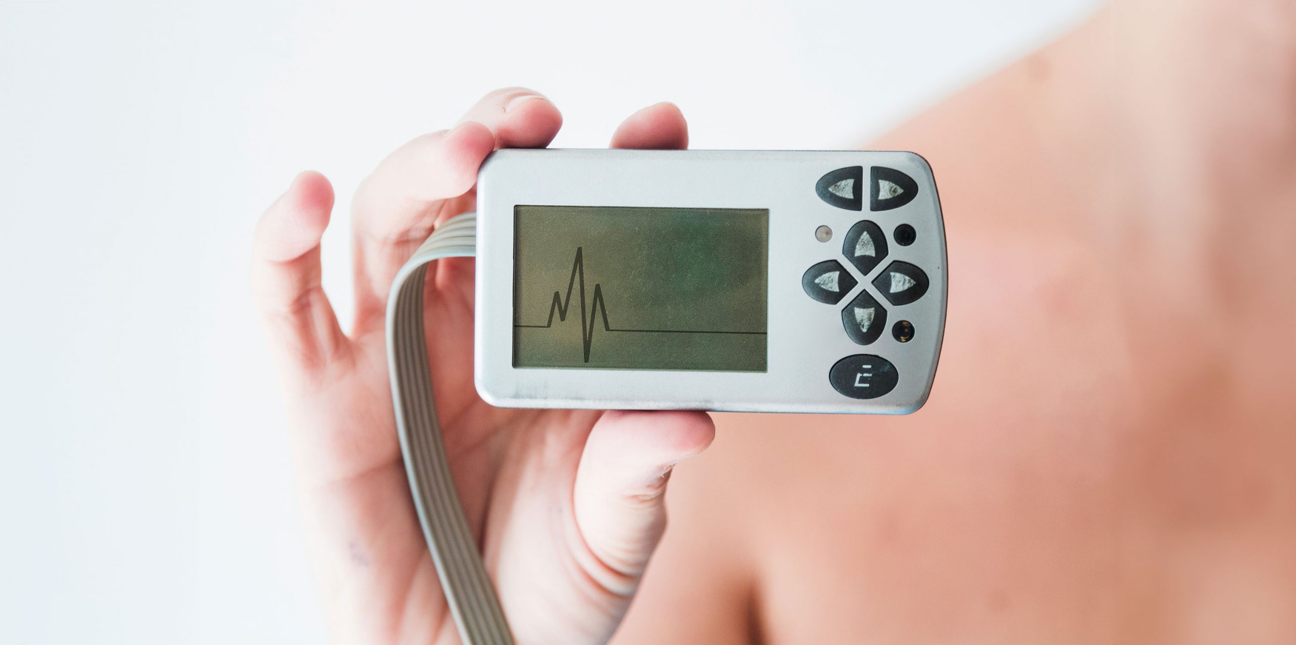
Enter the Holter monitor – a remarkable invention in the world of cardiac healthcare. Small in size but significant in impact, the Holter monitor serves as a vigilant guardian of heart health, offering insights that traditional methods might miss. It’s like having a miniature cardiac detective attached to you, meticulously recording every beat and rhythm of your heart. The data collected by this device is a treasure trove of information, providing a detailed picture of how your heart functions over an extended period, typically 24 to 48 hours.

Case Study: Holter Monitors: Revealing the Heart’s Secrets

Patient Information:

• Name: Sarah Thompson

• Background: Sarah, a 42-year-old office manager with no significant cardiovascular history, began experiencing occasional palpitations. Concerned about her heart health, she sought medical advice.

Early Challenges:

Sarah’s palpitations posed a diagnostic challenge as they occurred sporadically, making it difficult to capture irregularities during traditional ECG tests. The fleeting nature of her symptoms demanded a more prolonged monitoring solution.

Decision to Use Holter Monitor:

Recognizing the limitations of conventional testing, Sarah’s cardiologist recommended a 24-hour Holter monitor. This portable device offered continuous heart activity monitoring, capturing data throughout her daily activities and sleep, providing a comprehensive overview of her cardiac function.

Monitoring Strategies:

Sarah underwent comprehensive education on Holter monitor usage. In addition to wearing the device, she maintained a diary, logging daily activities and symptoms. This dual approach aimed to correlate symptoms with recorded data, enhancing the diagnostic value of the monitoring process.

Challenges During Monitoring:

While the Holter monitor was non-invasive, Sarah faced initial discomfort wearing the device. Additionally, consistently logging activities posed a challenge. However, these challenges were minor compared to the potential insights the monitor could provide.

End Results:

The 24-hour monitoring revealed episodes of arrhythmia during periods of increased stress at work. Detailed analysis by the healthcare team identified triggers for Sarah’s palpitations, leading to personalized treatment planning. Lifestyle modifications and stress management techniques were prescribed, resulting in a significant reduction in arrhythmia episodes during follow-up monitoring.

Statistical Improvement:

Before Holter monitoring, Sarah experienced palpitations 3-4 times a week, with high stress levels. After intervention, palpitations reduced to once a week, and her stress levels moderated, contributing to an improved overall quality of life.

Patient Feedback:

Sarah reported feeling more in control of her health and appreciated the non-invasive nature of the monitoring. She expressed gratitude for the personalized strategies that significantly improved her symptoms.
This case highlights the instrumental role of Holter monitors in diagnosing and managing cardiac issues with sporadic symptoms. Continuous monitoring, coupled with personalized interventions, led to a tangible improvement in the patient’s quality of life, emphasizing the importance of tailored approaches in cardiac care.

What is a Holter Monitor?

A Holter monitor is a sophisticated yet user-friendly device in the realm of cardiac care. This device comprises small electrodes, resembling plastic patches, that adhere to specific points on your chest and abdomen. These electrodes are connected to the monitor via wires, facilitating the continuous recording of your heart’s electrical activity. This method is non-invasive and painless, as no electricity is sent into the body.

Who Needs a Holter Monitor?

1. Symptoms Not Captured by Standard EKG: If a patient experiences symptoms like palpitations, chest pain, shortness of breath, skipped beats, or light-headedness, and these are not satisfactorily explained by a standard EKG, a Holter monitor may be recommended.

2. Unexplained Syncopal Episodes: Patients who have experienced unexplained fainting (syncope) or transient episodes of cerebral ischemia, which could potentially be related to cardiac rhythm disturbances, may benefit from Holter monitoring.

3. Cardiac or Conduction Disorders: Individuals diagnosed with significant cardiac or conduction disorders might require Holter monitoring as part of their evaluation and management. This continuous monitoring can provide detailed insights into the heart’s functioning over an extended period, helping in better diagnosis and treatment planning​​.

4. Heart Conditions with High Risk of Arrhythmias or Ischemia: For patients with heart conditions that are associated with a high incidence of serious cardiac arrhythmias or myocardial ischemia, a Holter monitor can be a crucial part of the evaluation. It helps in understanding the heart’s rhythm and its response to daily activities and stressors​​.

5. Evaluating the Effect of Cardiac Medications: If a patient is on cardiac medication that affects the electrical conduction system of the heart, Holter monitoring may be necessary to evaluate the effect of these medications on the patient’s cardiac rhythm and conduction system​​.

6. Pacemaker Functionality Checks: Patients with pacemakers who show clinical signs suggesting possible malfunction of the device may also be advised to undergo Holter monitoring. This is to ensure that the pacemaker is functioning correctly and managing the heart’s rhythm as intended​​.

Conditions Diagnosed by a Holter Monitor

A Holter monitor is a highly effective tool for diagnosing a variety of heart conditions, particularly those that may not be detected during a short-term ECG test. Here are some of the key conditions it helps identify:

1. Arrhythmias: Arrhythmias, or irregular heartbeats, are among the primary conditions detected by a Holter monitor. These include atrial fibrillation, bradycardia (slow heartbeat), tachycardia (fast heartbeat), and premature ventricular contractions (PVCs), which are extra, abnormal heartbeats originating in the ventricles​​​​.

2. Heart Palpitations and Unexplained Dizziness: Heart palpitations, often described as the heart racing or fluttering, and unexplained dizziness, are symptoms that can be linked to underlying heart rhythm issues. A Holter monitor helps in pinpointing these symptoms to specific heart rhythm abnormalities​​.

3. Evaluating Heart’s Oxygen Supply and Medication Effectiveness: The monitor also provides insights into how well the heart is getting oxygen and how effectively heart medications are working. This is crucial for patients with known heart conditions who are on medication​​.

4. Pacemaker Functionality: For patients with pacemakers, a Holter monitor can be used to assess how well the pacemaker is managing the heart’s rhythm​​.

5. Identifying Infrequent Arrhythmias: In cases where arrhythmias are infrequent, a standard ECG might not detect heart failure. A Holter Monitor Uses extend the monitoring period (24 to 72 hours or even longer), increasing the likelihood of capturing these sporadic events​​.

6. Further Diagnostic Steps: If a standard Holter monitor does not capture irregular heart rhythms, a healthcare provider might recommend an event recorder. This device, worn for a longer period (up to several weeks), allows patients to record their heart’s activity when they feel symptoms, providing further diagnostic information​​.

Comparing EKG and Holter Monitor

Function and Usage:

• Typically conducted in a doctor’s office, clinic, or hospital room, an EKG gives crucial information about the heart’s electrical activity. It shows if the electrical activity is normal, slow, fast, or irregular, and whether parts of the heart are enlarged or overworked​​.

• In contrast, a Holter monitor is a portable device used for continuous ECG monitoring over several days. It is particularly useful for detecting cardiac events such as atrial fibrillation and suspected stroke that may not be captured during the brief period of an EKG​​.

Recording Duration:

• The EKG provides a snapshot of the heart’s activity at a single point in time. It is extremely valuable for diagnosing arrhythmias but is limited to capturing heart activity only during the test. This poses a challenge for intermittent arrhythmias that may not occur during the EKG​​.

• The Holter monitor, however, records heart activity continuously, typically for 24 to 48 hours, and sometimes longer. This feature allows it to capture intermittent arrhythmias and correlate them with the patient’s symptoms. Even asymptomatic episodes are recorded, providing a comprehensive picture of heart activity​​.

Scope of Monitoring:

• The EKG is an essential, noninvasive diagnostic tool for many common heart problems in people of all ages. However, it may not always provide a comprehensive result, especially for conditions that manifest intermittently​​.

• The Holter monitor offers a more complete picture of heart activity, necessary for the diagnosis of certain cardiac conditions. Its continuous recording is more likely to detect irregularities that might be missed by an EKG​​.

Technological Advancements and Alternatives

• Apart from traditional EKGs and Holter monitors, there are newer technologies like smartwatches and insertable cardiac monitor uses. For instance, the Apple Watch can screen for asymptomatic events and record ECG rhythm strips on demand. However, they have limitations like false positive detections and require careful usage​​.

• Insertable cardiac monitors provide very long-term monitoring, up to three years or so. They can detect significant arrhythmias and save recordings automatically, but they require an invasive procedure for insertion and removal​​.

Procedure of Wearing a Holter Monitor

Initial Setup

• A technician attaches the monitor and provides instructions. The setup involves attaching electrodes to the chest. These electrodes are small, round, sticky patches that record the heart’s electrical activity. In some cases, the technician may need to shave areas of the chest to ensure the electrodes attach firmly​​.

Placing the Monitor

• The technician assists in putting on the monitor and connecting it to the electrodes. They provide guidance on how to care for the monitor. The monitor is typically carried in a pocket or bag, or worn on a strap like a purse or on the waist​​.

Daily Activities and Restrictions

• Patients can engage in most of their usual activities while wearing the monitor. However, they should avoid bathing, showering, swimming, undergoing X-rays, and being near high-voltage areas or metal detectors to prevent damage to the device or interference with its functioning​​.

Activity and Symptom Diary

• The wearer is advised to keep a diary of activities and any symptoms experienced, such as shortness of breath, chest pain, or irregular heartbeats. This diary helps correlate any symptoms with changes in the heart’s electrical activity recorded by the Holter monitor​​.

Post-Monitoring Process

• After wearing the monitor for the prescribed 24- or 48-hour period, it is returned to the technician. The technician then processes the records, including the EKG data and the patient’s notes, and prepares a report for the healthcare provider.

Conclusion

In conclusion, the Holter monitor is a pivotal tool in cardiac health, providing detailed and continuous analysis of heart rhythms over an extended period. Its ability to detect and characterize various arrhythmias, assess the effectiveness of medications, and identify potential cardiac conditions is invaluable. The correlation of arrhythmias with patient symptoms offers a deeper understanding of each individual’s cardiac health, enabling personalized medical interventions.

Based on the insights provided in this blog about the Holter monitor and its significance in cardiac health, Dr. Ellen Mellow, with her expertise in cardiology, can offer personalized care and detailed analysis of Holter monitor results. Her knowledge and experience in interpreting these results can be crucial in diagnosing and managing various heart conditions effectively, ensuring patients receive the most informed and tailored cardiac care possible.

Conventional and Sophisticated Medical Diagnostic Procedures for Heart Problems

Introduction

Heart and cardiovascular diseases are serious health issues that cause many deaths around the world. Detecting these problems early is very important for the success of treatment and management. Thankfully, there are many advanced medical tests available today that can detect heart problems effectively. In this blog post, we will discuss some of the most commonly used and advanced tests that doctors use to diagnose heart issues. We’ll explain why these tests are important and how they work.

ECG – electrocardiogram

Heart and cardiovascular diseases are some of the most common causes of death across the world. Detecting them early is crucial for effective treatment and management. Fortunately, modern science has come up with several tests that can detect heart problems accurately. In this blog post, we’ll take a look at some of the most sophisticated and widely used medical tests for diagnosing cardiac issues. We’ll explain why they are important and how they work.

Echocardiogram

Using sound waves, an echocardiogram produces images of your heart. This examination is crucial for assessing the architecture and physiology of the heart. During the procedure, a transducer is a device that is placed on your chest. It records the echoes that return after sending sound waves to the heart. These pictures can show conditions such as unusual heart size, malfunctioning cardiac valves, and poor heart muscle activity.

Stress Examination

This procedure, commonly called an exercise stress test, evaluates how effectively your heart works during physical activity. Exercise-induced increases in heart rate and force may be a sign of cardiac problems that are not evident when the heart is at rest. During a stress test, your heart rate will be monitored as you walk on a treadmill or ride on a stationary bike.
If you are unable to exercise, you may be administered medication that simulates the heart-related benefits of exercise.

Cardiac Catheterization

The cardiac catheterization procedure is a more invasive diagnostic technique. A thin, flexible tube called a catheter is inserted into a blood vessel, usually in your arm or groin, and guided into your heart. With this catheter, doctors may perform diagnostic tests and provide medications. They can measure arterial blockages, draw blood samples, and keep an eye on the pressure inside the heart’s chambers. This test is necessary for the detection of coronary artery disease as well as other heart conditions.

Blood Tests

When it comes to identifying cardiac issues, blood testing is essential. Blood tests for specific enzymes and proteins may reveal damage to the heart muscle. For instance, elevated levels of troponin, a protein produced in response to myocardial injury, are frequently indicative of myocardial infarction. Additionally, blood testing can evaluate additional heart disease risk factors including cholesterol levels.

Advanced Medical Diagnostics for Heart Problems

Diagnostic technology has advanced significantly in the field of cardiology. These advanced diagnostic procedures offer a more profound understanding of the health and function of the heart, enabling early and precise identification of cardiac disorders.

Cardiac MRI (Magnetic Resonance Imaging)

A very sophisticated imaging technique called cardiac MRI is utilized to provide finely detailed images of the heart. A cardiac MRI, in contrast to a general MRI, concentrates on the heart and the tissues around it. Heart tumors, congenital heart abnormalities, heart muscle illnesses, and the consequences of coronary artery disease can all be detected with this test. Without subjecting the patient to radiation, it offers thorough information on the composition and operation of the heart.

CT Coronary Angiogram

A non-invasive imaging procedure called a CT coronary angiography aids in the visualization of the arteries that deliver blood to the heart. To create high-resolution pictures of the coronary arteries, a computed tomography (CT) scanner and an iodine-containing contrast dye are used. Plaque buildup and heart attack risk can be assessed with this test. The procedure is especially beneficial to patients who find it difficult to undergo traditional angiograms.

Positron Emission Tomography (PET) Scan

A PET scan is a high-tech imaging procedure that shows the heart’s metabolic activity by using a little quantity of radioactive material. Particularly in the aftermath of a heart attack, it might offer details on blood flow and the health of the heart muscle. PET scans are frequently used in conjunction with other diagnostic procedures to evaluate the effects of a heart attack and detect coronary artery disease.

Intravascular Ultrasound (IVUS)

During a cardiac catheterization, intravascular ultrasound is a sophisticated operation. A small ultrasonography probe is put into the coronary arteries by means of a catheter tip. It offers an insider’s perspective of the arterial walls in great detail. When considering procedures such as the installation of stents, IVUS is very helpful in determining the degree of plaque formation.

Tilt Table Test

The purpose of the tilt table test is to identify the reasons for syncope, or fainting. The patient is strapped to a table during the test, and the table is tilted at different angles to measure the patient’s heart rate, blood pressure, and other physiological reactions. This test aids in the diagnosis of disorders related to the heart’s incapacity to react appropriately to changes in body posture, such as orthostatic hypotension and vasovagal syncope.

The Role of Technology in Advancing Cardiac Diagnostics

Technology has completely changed the field of cardiac diagnostics, enabling more accurate, non-invasive, and educational testing. The merger of several technical domains has resulted in this progress.

Imaging Technology

Technological developments in imaging have been essential. The anatomy and function of the heart may now be seen in previously unheard-of detail thanks to high-resolution imaging methods like CT and MRI scans. Not only have pictures become clearer, but they are also captured faster, which is important for an organ like the heart that is always in motion. Examples of how technology has improved our capacity to examine the heart non-invasively and with high precision include cardiac MRIs and CT coronary angiograms.

Software and Data Analysis

The emergence of advanced software for analyzing and interpreting data has had a revolutionary effect. Complex cardiac data analysis is increasingly being done with the help of artificial intelligence (AI) and sophisticated algorithms. In addition to enhancing the accuracy of diagnoses, this software can detect patterns and anomalies that humans might miss. An example of how software innovation supports cardiac care is the integration of artificial intelligence into the interpretation of PET scans and ECG readings.

Miniaturization and Material Science

Procedures like Intravascular Ultrasound (IVUS) are now possible due to the downsizing of medical instruments brought about by advances in material science. Cardiologists now get a closer, more precise look at the inside components of the heart thanks to the integration of small ultrasound probes on catheters. Additionally, by being smaller, cardiac devices are now less intrusive and more pleasant for patients.

Wireless and Wearable Technologies

Continuous cardiac monitoring now provides more options because of the advancement of wearable and wireless technology. Heart health may be tracked in real time using gadgets like smartwatches and portable ECG monitors, which offer useful information outside of the professional context. For the early diagnosis and treatment of chronic cardiac diseases, this ongoing monitoring is essential.

Telemedicine and Remote Monitoring

Last but not least, the development of telemedicine and remote monitoring tools has altered the provision of cardiac care. The treatment of chronic cardiac illnesses and post-operative care can now be improved by patients sharing real-time health data with their doctors from the comfort of their own homes. With the use of this technology, patients can receive ongoing treatment with fewer frequent hospital visits.

Conclusion

The timely identification of cardiac disorders is essential for efficient therapy and can greatly enhance results. Crucial information on the health of the heart may be obtained from these tests, which range from non-invasive ECGs and echocardiograms to more involved procedures like cardiac catheterization. It’s crucial to speak with a medical expert if you have concerns about the health of your heart. They can recommend the right tests for your circumstances. Recall that prompt cardiac treatment can prevent fatalities.

Dr. Mellow provides individualized cardiac and preventive care solutions based on his more than 24 years of expertise in cardiology and general medicine. Her business is renowned for utilizing cutting-edge cardiac technology, which enables her to offer electronic treatment to patients all around the world. As a member of New York-Presbyterian Hospital/Weill Cornell Medicine, Dr. Mellow provides sophisticated and comprehensive cardiac care to her patients.

What Is Preventive Health and Why Is It Important?

Introduction

Imagine taking control of your health, not just when you’re ill, but every day. This is what preventive health is all about. It’s a proactive approach, like nurturing a garden, ensuring it flourishes and remains free from weeds. Instead of waiting for symptoms to show up, preventive health encourages regular care and maintenance of your body and mind. It’s about understanding the small, everyday choices that can significantly impact your well-being in the long run.

From the foods you eat to the walks you take, and the regular health checks you shouldn’t skip – each of these acts as a building block for a healthier, more vibrant you. In essence, preventive health isn’t just a medical concept; it’s a way of living that puts you in the driver’s seat of your health journey.

The Basics of Preventive Health

Preventive health is a proactive approach that puts patients first. It’s about preventing health issues before they occur, rather than waiting until they arise. There are three tiers of preventive health: primary, secondary, and tertiary. Primary prevention involves preventing diseases before they occur, like getting vaccinated. Secondary prevention is about detecting health problems early, such as routine screenings for cancer. Tertiary prevention aims to reduce the impact of existing diseases. Preventive health empowers patients with knowledge, tools, and guidance to take charge of their well-being. It’s a vital tool on the path to a healthier, happier life.

Why Preventive Health Matters to You

Early Disease Detection and Prevention:

Early detection of diseases, particularly cancer, has a significant impact on survival rates. For example, the survival rate for women with early-detected cervical cancer is remarkably higher compared to when it’s diagnosed at a later stage. This pattern is consistent across various types of cancer, where early detection drastically improves outcomes. Similarly, for chronic conditions like heart disease and diabetes, early diagnosis can prevent complications and improve overall health. It’s important to highlight how regular screenings and being aware of changes in one’s health can lead to early detection, which in many cases, can be life-saving.

Reducing Medical Expenses:

Early detection and management of health conditions can significantly reduce medical expenses in the long run. By catching diseases early, patients can avoid costly treatments and hospital stays associated with advanced stages of diseases. This is particularly true for chronic conditions like diabetes and heart disease, where early intervention can prevent complications that require more intensive and expensive care.

Improving Quality of Life:

Preventive health not only extends life expectancy but also enhances the quality of life. Managing chronic conditions effectively and maintaining a healthy lifestyle can lead to better mental and physical health, more energy, and a reduced risk of complications. The idea here is to emphasize that preventive health is not just about living longer but living better.

Empowerment Through Knowledge:

Educating oneself about health risks and how to manage them is a form of empowerment. Encourage your readers to be proactive about their health, seeking regular check-ups, and staying informed about their health conditions. This section can emphasize the importance of a partnership with healthcare providers, where patients are active participants in their health care.

By focusing on these areas, your blog can effectively communicate the importance of preventive health to a B2C audience, emphasizing the practical and personal benefits. Remember to maintain a simple and engaging tone, making the content relatable and easy to understand for patients. The goal is to inspire readers to take charge of their health proactively, highlighting how preventive measures are not just medical procedures but integral parts of a healthier lifestyle.

Key Preventive Health Practices for Patients

Routine Health Screenings and Check-Ups:

Regular medical check-ups are vital for early detection and effective treatment of various health conditions, including chronic illnesses such as hypertension and diabetes, and diseases like cancer. For instance, screenings like Pap tests for cervical cancer and mammograms for breast cancer can detect these conditions early when the chance of cure is high. Similarly, colorectal and lung cancer screenings play a crucial role in finding these cancers at a stage when they are more treatable. Emphasize to your readers that these screenings are essential even when they feel healthy, as many conditions do not show symptoms in the early stages.

Monitoring Existing Conditions:

For patients with diagnosed medical conditions, regular check-ups are key in monitoring the progression of these diseases. These check-ups help assess the effectiveness of ongoing treatments and make necessary adjustments. They are essential for managing chronic diseases effectively, reducing the risk of complications, and improving the overall quality of life.

Lifestyle Consultations:

During check-ups, healthcare providers often offer advice on improving lifestyle, which is integral to long-term well-being. This includes dietary recommendations, physical activity guidelines, and stress management strategies. Tailored advice based on individual health needs can significantly impact overall health.

Vaccination:

Up-to-date vaccinations play a critical role in maintaining immunity and protecting against infectious diseases. They are important at all stages of life, from childhood to adulthood. Vaccinations not only protect the individual but also contribute to community health by creating herd immunity.

Aging Management:

As we age, our health needs change. Regular medical examinations for older adults are crucial for detecting age-related health changes and managing chronic diseases effectively. They help in monitoring risk factors like high blood pressure, cholesterol levels, and diabetes, and in maintaining functional abilities and psychological well-being.

Ensure to communicate these points in a simple, easy-to-understand manner, focusing on how these practices directly benefit the patient. The goal is to encourage readers to proactively manage their health through these preventive practices, emphasizing the long-term benefits of regular health check-ups and lifestyle modifications.

The connection between preventive health and cardiovascular wellness

The connection between preventive health and cardiovascular wellness is a critical aspect of modern healthcare, especially considering cardiovascular disease (CVD) is the leading cause of death globally. Despite advancements in treatment reducing mortality rates, the global burden of CVD remains high, largely due to its preventable nature. This context underscores the importance of focusing on cardiovascular wellness as part of preventive health.

Preventing atherosclerotic cardiovascular disease (ASCVD) means promoting “health factors” to cultivate cardiovascular wellness. This approach understands that cardiovascular health is more than just the absence of disease. It requires a proactive stance towards health, treating risk factors and encouraging positive health behaviors.

Healthy lifestyle (HL) behaviors can prevent CVD. These include regular exercise, healthy eating habits, adequate sleep, and quitting smoking. Such habits affect traditional CVD risk factors and cardiorespiratory fitness (CRF), which is a significant but less commonly measured risk factor. Cardiac rehabilitation programs focus on secondary prevention, but also include comprehensive HL behavior changes. By adopting a comprehensive approach to lifestyle modification, we can improve CRF and other risk factors, reducing the burden of CVD.

Clinicians play a crucial role in promoting HL behaviors to prevent CVD. They are in a unique position to educate and motivate patients about the importance of lifestyle changes as a means of CVD prevention. This involves not only providing information but also supporting patients in making sustainable lifestyle changes.

To keep your heart healthy, it’s important to focus on preventing health issues by promoting healthy habits and factors that go beyond simply addressing disease risks. The current approach to preventing cardiovascular diseases is comprehensive, combining medical treatments with lifestyle changes to promote overall cardiovascular wellbeing. By combining both approaches, we can better protect our hearts and overall health.

Conclusion

As we wrap up our exploration of preventive health, it’s clear that being proactive about our health is not just a choice but a necessity. Preventive health is about more than avoiding illness – it’s about embracing a lifestyle that fosters longevity, vitality, and well-being. By understanding the importance of regular health screenings, vaccinations, maintaining a healthy lifestyle, and being mindful of our mental health, we empower ourselves to live our best lives.

It’s important to recognize that while there are barriers to accessing preventive health, from financial constraints to cultural misconceptions, these challenges are not insurmountable. With the right information, support, and motivation, each of us can navigate these hurdles. We’re not just patients in a healthcare system; we’re active participants in our journey towards health and wellness.

Dr. Ellen Mellow, a highly experienced cardiologist and internist in New York, provides personalized and advanced cardiac and preventive care, catering to a wide range of patient needs both locally and globally through technology

5 warning signs of a heart attack that you may not know

Introduction

Imagine this: You’re going about your day, busy with life’s usual tasks, when suddenly you feel something unusual. It’s not the dramatic chest clutching we often see in movies. Instead, it’s something subtler, maybe even something you’d brush off as just stress or fatigue. But what if these seemingly minor signs were actually warning bells of a heart attack?

Heart attacks are often thought of as dramatic and obvious, but sometimes, they sneak up quietly, presenting symptoms that are easy to miss or misinterpret. In this blog, we’re going to shed light on five such warnings of heart attack that might not be on everyone’s radar.

1. Pain in Arm, Jaw, Neck, or Back

Sometimes, the warning signs of a heart attack are not where you’d expect. Pain in areas like the arm, jaw, neck, or back can be subtle messengers. These pains might feel different from typical muscle aches – they could be persistent, sudden, or even come and go. This type of discomfort is especially noteworthy if it’s unusual for you. It’s not just about severity; it’s about the peculiarity of this pain. It’s your body’s way of sounding an alarm that something might not be right with your heart.

• Look out for unusual, persistent, or intermittent pain in these areas.

• Pain may not always be severe but pay attention to its unusual nature.

• Especially important to note if these pains are not typical for you.

2. Cold Sweats:

Imagine you’re not exerting yourself, but suddenly, you find yourself in a cold sweat. This isn’t your typical response to a hot day or physical activity; it’s different. It’s as if your body is reacting to something internally. Cold sweats can be a sneaky cause of heart failure, often overlooked because they seem so benign. But when coupled with other signs, they shouldn’t be ignored. It’s like your body’s cooling system is kicking in without any apparent reason.

• Sudden onset of cold sweats without physical exertion is a red flag.

• Different from normal sweating, may feel more intense or unexplained.

• Pay attention when coupled with other subtle symptoms.

3. Heartburn or Indigestion:

That discomfort you’re dismissing as just a bit of heartburn or a reaction to your last meal? It might be more than that. Sometimes, what feels like a simple case of indigestion or heartburn could actually be a heart attack in disguise. The tricky part is distinguishing between usual digestive issues and heart-related pain. If this ‘heartburn’ feels different, more severe, or occurs in conjunction with other symptoms, it’s time to take it seriously.

• Heartburn-like pain may be a heart attack sign, especially if it’s atypical for you.

• Severity and timing can help differentiate it from normal indigestion.

• Consider other simultaneous symptoms for a clearer picture.

4. Shortness of Breath:

You’re not running a marathon, yet you find yourself short of breath. This unexplained, sudden difficulty in breathing can be more than just a lack of fitness or a momentary discomfort. It could be your heart telling you that it’s struggling. When your heart can’t pump effectively, your lungs don’t get enough oxygen, leading to this frightening experience of gasping for air. It’s a silent but urgent call for attention.

• Unexplained difficulty in breathing, especially when resting, could be critical.

• The heart and lungs work together – issues with one can affect the other.

• Take note if this symptom occurs suddenly or feels out of the ordinary.

5. Nausea or Vomiting

Nausea or sudden vomiting can be easily mistaken for a stomach bug or food poisoning, but sometimes, the cause lies deeper. These symptoms can be unexpected heralds of a heart attack, particularly in women. It’s surprising how a symptom so commonly associated with the digestive system can be linked to the heart. But when your heart is in distress, your whole body reacts in ways you might not anticipate.

• Sudden nausea or vomiting without a clear cause should be monitored closely.

• Particularly important for women, as they often have atypical heart attack symptoms.

• Consider the context and other symptoms for a comprehensive understanding.

Difference in Symptoms Between Men and Women

When it comes to heart attacks, the symptoms can vary significantly between men and women, leading to a dangerous gap in awareness and timely response. It’s a common misconception that heart attack symptoms are uniform across genders. However, research and clinical observations suggest that women often experience different, subtler symptoms compared to men.
For men, the classic symptom of a heart attack is chest pain or discomfort, which is also common in women. However, women are more likely to experience symptoms that are less traditionally associated with heart attacks. These can include shortness of breath, nausea or vomiting, and back or jaw pain. In fact, some women may have a heart attack without experiencing any chest pain at all.

It’s not just the type of symptoms that differ, but also their presentation. Women’s symptoms can be more vague and less intense than the stereotypical heart attack symptoms. For example, rather than experiencing crushing chest pain, a woman might feel a squeezing sensation or a sense of fullness in the chest. Shortness of breath in women might not be accompanied by chest pain, which can lead to misdiagnosis or delay in seeking treatment.
Furthermore, women might experience these symptoms more often when resting, or even while asleep, unlike men who might notice symptoms more during physical exertion. Unusual fatigue, lightheadedness, and indigestion-like feelings are also more prevalent as silent heart attack signs in women.

Understanding these differences is crucial, as they can impact how quickly individuals recognize heart attack symptoms and seek medical attention. Women, in particular, should be aware of these atypical symptoms and take them seriously. Timely medical intervention is key in treating heart attacks, and knowing these gender-specific symptoms can save lives.

What to Do If You Suspect a Heart Attack

If you or someone around you exhibits any of the discussed warning signs of a heart attack, the first and most crucial step is to act immediately. Heart attacks are medical emergencies where every minute counts, and prompt action can save a life.

The initial action should always be to call emergency services. In many countries, this would be dialing 911 or the local equivalent. It’s essential to understand that a heart attack is a time-sensitive emergency. The longer it takes to receive treatment, the more damage the heart muscle sustains. Emergency responders are trained to deliver life-saving care en route to the hospital, which can be crucial in managing a heart attack.

Conclusion

Heart attacks, as we’ve seen, don’t always announce themselves with clear, unmistakable signs. Often, they manifest through subtle symptoms that are easy to overlook or misinterpret. This underscores the importance of listening to our bodies and being vigilant about changes, no matter how minor they may seem.

It’s crucial to remember that heart attacks can happen to anyone, regardless of age, gender, or perceived health status. Understanding the variety of symptoms, especially those that are more prevalent in women, can be lifesaving knowledge. Equally important is knowing what to do if you or someone else exhibits these signs – acting swiftly, calling for emergency help, and administering first aid if necessary.

Dr. Ellen Mellow, one of the best heart specialists in NYC, with over 24 years of experience in cardiology and internal medicine, provides expert care in various aspects of cardiac health. Her practice focuses on personalized cardiac and preventive care solutions, addressing complex health conditions through a holistic approach. This includes advanced lipid profiling, electrocardiography (ECG/EKG), Holter monitoring, pre-operative cardiac clearance, and management of chronic disorders. Dr. Mellow’s comprehensive assessments aid in diagnosing and treating intricate cardiac conditions, emphasizing patient-focused care.

Abdominal Aortic Doppler: What to Expect Before, During, and After

Introduction

The abdominal aortic Doppler ultrasound stands as a critical diagnostic tool in modern medicine. This guide aims to demystify each stage of the procedure, providing in-depth insights from preparation to recovery. Tailored for patients and caregivers, it’s designed to inform, alleviate concerns, and enhance understanding of this vital medical examination.

Understanding the Function of the Abdominal Aorta

The principal blood artery that provides blood to the lower body is the abdominal aorta. Aneurysms and blockages are examples of disorders of this important artery that can cause major health problems. To find these problems, an abdominal aortic Doppler ultrasonography is essential. This non-invasive technique helps medical professionals spot anomalies by using sound waves to make photographs of the blood flow via the aorta. This Doppler’s early identification is essential for prompt medical action, which has the ability to save lives and avoid consequences.

• Main vessel for lower body blood supply.

• Detects conditions like aneurysms and blockages.

• Non-invasive, uses sound waves for imaging.

• Early detection is crucial for effective treatment.

Preparing for an Abdominal Aortic Doppler

In order to guarantee reliable findings, there are a few crucial procedures to prepare for an abdominal aortic Doppler. In order to cleanse the stomach and intestines for the screening process, patients are frequently needed to fast for a few hours beforehand. This is because food and liquids might obstruct the abdominal ultrasound pictures. Furthermore, it’s critical to review existing drugs with the doctor because some may require adjustments. Being aware of the process in advance can assist reduce anxiety and improve comfort levels. It’s essential to follow these preparatory procedures for a seamless and successful Doppler ultrasonography.

• Fasting is required to clear the stomach and intestines.

• Discuss medication adjustments with the doctor.

• Understanding the procedure helps reduce anxiety.

• Preparation is key for effective results.

The Procedure: During the Abdominal Aortic Doppler

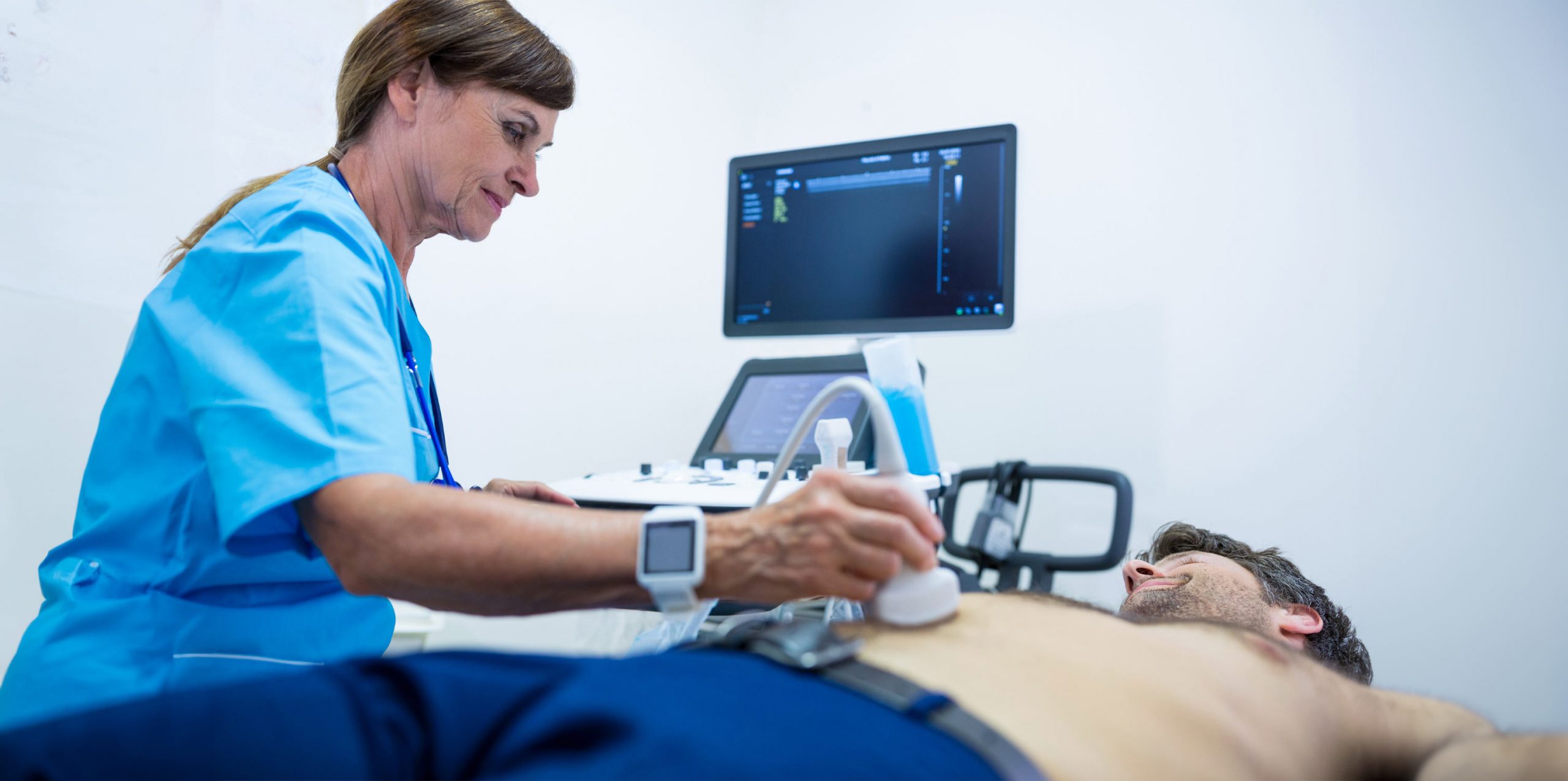
Patients recline on an examination table while a special gel is placed to their abdomens for the abdominal aortic Doppler. The sound waves are better transmitted by this gel. The sonographer then transmits and receives these waves using a transducer to produce pictures of the aorta on a monitor. Though some people may experience some little discomfort due to the transducer’s pressure, the procedure is usually pleasant. In less than an hour, the treatment usually yields useful information regarding the abdominal aorta test and health.

• Patients lie on a table, gel applied to the abdomen.

• Transducers used to send/receive sound waves.

• Generally painless, slight discomfort possible.

• Takes less than an hour, and provides crucial information.

Interpreting the Results

The results of an abdominal aortic Doppler are interpreted by a radiologist. Normal results show a consistent size and shape of the aorta with no signs of bulging or abnormal blood flow patterns. Abnormal results may indicate issues like aneurysms or blockages. However, these findings don’t always signify a severe condition and may require further testing for accurate diagnosis. The radiologist’s expertise is key in interpreting these images, providing essential information for the patient’s ongoing medical care.

• Radiologist interprets the results.

• Normal results show consistent aorta size and shape.

• Abnormal results may indicate aneurysms or blockages.

• Further testing may be required for accurate diagnosis.

After the Procedure: Recovery and Follow-up

Recovery from an abdominal aortic Doppler is typically straightforward. Even if uncommon symptoms and pain are uncommon, it’s crucial to let the doctor know about them. In light of the findings, the physician can advise routine observation to monitor any alterations in the state of the aorta. In order to manage any health risks and guarantee the best possible results, this follow-up is essential.

• Normal activities can be resumed immediately.

• Report any unusual symptoms to the doctor.

• Regular monitoring may be recommended.

• Follow-up is crucial for ongoing health management.

Risks and Complications

The abdominal aortic Doppler is a safe procedure with minimal risks. The most common issues patients might experience are minor discomfort or skin irritation from the gel used during the ultrasound. Major side effects are quite uncommon because the test is non-invasive. Still, being a knowledgeable patient requires being aware of these possible hazards, no matter how slight.

• Procedure is safe with minimal risks.

• Common issues: minor discomfort, skin irritation.

• Major complications are extremely rare.

• Awareness of potential risks is important.

Advances in Technology: The Role of Abdominal Aorta Ultrasound

The realm of abdominal aorta ultrasound represents a significant technological advancement in cardiovascular diagnostics. This state-of-the-art tool leverages high-frequency sound waves to generate real-time images of the abdominal aorta, providing crucial insights into its structure and function. The sophistication of this technology lies in its ability to detect minute changes in the aorta, such as the early formation of aneurysms or the presence of blockages, with remarkable precision. Continuous advancements in ultrasound technology, including enhanced image clarity and non-invasive nature, make it an indispensable tool in modern cardiology, aiding in early diagnosis and timely intervention.

• Utilizes high-frequency sound waves for imaging.

• Detects minute changes in the abdominal aorta.

• Continuous advancements enhance image clarity.

• Non-invasive and critical for early diagnosis.

Conclusion

In summary, the abdominal aortic Doppler ultrasonography provides a comprehensive view of the condition of the abdominal aorta, making it an essential tool for cardiovascular health. We have made an effort to explain every aspect of the process in this guide, from the preliminary planning to the careful analysis of the findings and the follow-up.

Ellen Mellow, MD, stands out as a superb option for cardiology and primary care by drawing on the thorough insights offered in our guide on the abdominal aortic Doppler ultrasound. Being a member of the American College of Cardiology and the American Heart Association, Dr. Mellow’s knowledge is especially helpful in assisting patients in understanding the complexities of cardiovascular health, guaranteeing comprehensive treatment and well-informed choices when undergoing procedures such as abdominal aortic Doppler.