Our Promise
Trustworthy Medical Care
Get your care delivered by a board-certified physician with an experience of over 24 years
Book Your Appointment
Whenever you need to, simply call us or book your appointment online
The abdominal aortic Doppler ultrasound stands as a critical diagnostic tool in modern medicine. This guide aims to demystify each stage of the procedure, providing in-depth insights from preparation to recovery. Tailored for patients and caregivers, it’s designed to inform, alleviate concerns, and enhance understanding of this vital medical examination.
The principal blood artery that provides blood to the lower body is the abdominal aorta. Aneurysms and blockages are examples of disorders of this important artery that can cause major health problems. To find these problems, an abdominal aortic Doppler ultrasonography is essential. This non-invasive technique helps medical professionals spot anomalies by using sound waves to make photographs of the blood flow via the aorta. This Doppler’s early identification is essential for prompt medical action, which has the ability to save lives and avoid consequences.
• Main vessel for lower body blood supply.
• Detects conditions like aneurysms and blockages.
• Non-invasive, uses sound waves for imaging.
• Early detection is crucial for effective treatment.
In order to guarantee reliable findings, there are a few crucial procedures to prepare for an abdominal aortic Doppler. In order to cleanse the stomach and intestines for the screening process, patients are frequently needed to fast for a few hours beforehand. This is because food and liquids might obstruct the abdominal ultrasound pictures. Furthermore, it’s critical to review existing drugs with the doctor because some may require adjustments. Being aware of the process in advance can assist reduce anxiety and improve comfort levels. It’s essential to follow these preparatory procedures for a seamless and successful Doppler ultrasonography.
• Fasting is required to clear the stomach and intestines.
• Discuss medication adjustments with the doctor.
• Understanding the procedure helps reduce anxiety.
• Preparation is key for effective results.
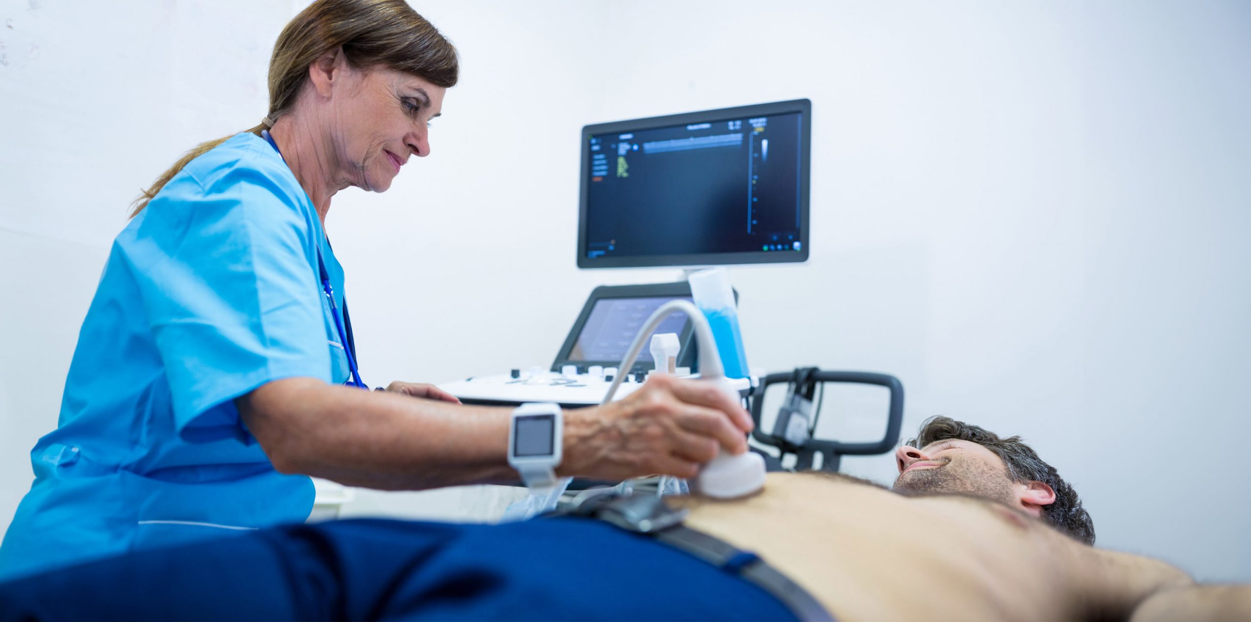
Patients recline on an examination table while a special gel is placed to their abdomens for the abdominal aortic Doppler. The sound waves are better transmitted by this gel. The sonographer then transmits and receives these waves using a transducer to produce pictures of the aorta on a monitor. Though some people may experience some little discomfort due to the transducer’s pressure, the procedure is usually pleasant. In less than an hour, the treatment usually yields useful information regarding the abdominal aorta test and health.
• Patients lie on a table, gel applied to the abdomen.
• Transducers used to send/receive sound waves.
• Generally painless, slight discomfort possible.
• Takes less than an hour, and provides crucial information.
The results of an abdominal aortic Doppler are interpreted by a radiologist. Normal results show a consistent size and shape of the aorta with no signs of bulging or abnormal blood flow patterns. Abnormal results may indicate issues like aneurysms or blockages. However, these findings don’t always signify a severe condition and may require further testing for accurate diagnosis. The radiologist’s expertise is key in interpreting these images, providing essential information for the patient’s ongoing medical care.
• Radiologist interprets the results.
• Normal results show consistent aorta size and shape.
• Abnormal results may indicate aneurysms or blockages.
• Further testing may be required for accurate diagnosis.
Recovery from an abdominal aortic Doppler is typically straightforward. Even if uncommon symptoms and pain are uncommon, it’s crucial to let the doctor know about them. In light of the findings, the physician can advise routine observation to monitor any alterations in the state of the aorta. In order to manage any health risks and guarantee the best possible results, this follow-up is essential.
• Normal activities can be resumed immediately.
• Report any unusual symptoms to the doctor.
• Regular monitoring may be recommended.
• Follow-up is crucial for ongoing health management.
The abdominal aortic Doppler is a safe procedure with minimal risks. The most common issues patients might experience are minor discomfort or skin irritation from the gel used during the ultrasound. Major side effects are quite uncommon because the test is non-invasive. Still, being a knowledgeable patient requires being aware of these possible hazards, no matter how slight.
• Procedure is safe with minimal risks.
• Common issues: minor discomfort, skin irritation.
• Major complications are extremely rare.
• Awareness of potential risks is important.
The realm of abdominal aorta ultrasound represents a significant technological advancement in cardiovascular diagnostics. This state-of-the-art tool leverages high-frequency sound waves to generate real-time images of the abdominal aorta, providing crucial insights into its structure and function. The sophistication of this technology lies in its ability to detect minute changes in the aorta, such as the early formation of aneurysms or the presence of blockages, with remarkable precision. Continuous advancements in ultrasound technology, including enhanced image clarity and non-invasive nature, make it an indispensable tool in modern cardiology, aiding in early diagnosis and timely intervention.
• Utilizes high-frequency sound waves for imaging.
• Detects minute changes in the abdominal aorta.
• Continuous advancements enhance image clarity.
• Non-invasive and critical for early diagnosis.
In summary, the abdominal aortic Doppler ultrasonography provides a comprehensive view of the condition of the abdominal aorta, making it an essential tool for cardiovascular health. We have made an effort to explain every aspect of the process in this guide, from the preliminary planning to the careful analysis of the findings and the follow-up.
Ellen Mellow, MD, stands out as a superb option for cardiology and primary care by drawing on the thorough insights offered in our guide on the abdominal aortic Doppler ultrasound. Being a member of the American College of Cardiology and the American Heart Association, Dr. Mellow’s knowledge is especially helpful in assisting patients in understanding the complexities of cardiovascular health, guaranteeing comprehensive treatment and well-informed choices when undergoing procedures such as abdominal aortic Doppler.
Get your care delivered by a board-certified physician with an experience of over 24 years
Whenever you need to, simply call us or book your appointment online